Squamous Cell Carcinoma Base Of Tongue Radiology
Squamous Cell Carcinoma Tongue Radiology Reference Article Radiopaedia Org

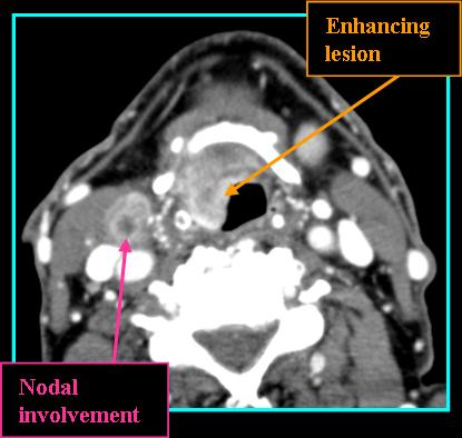
Base Of Tongue Tumor Squamous Cell Carcinoma Radiology Case Radiopaedia Org

Base Of Tongue Squamous Cell Carcinoma Radiology Case Radiopaedia Org
Tongue Squamous Cell Carcinoma Crossing Midline Radiology Case Radiopaedia Org

Squamous Cell Carcinoma Of The Tongue Radiology Case Radiopaedia Org
Tongue And Floor Of Mouth Neoplasm Radiology Case Radiopaedia Org

Scca comprises over 95 of all malignancies of the tongue.
Squamous cell carcinoma base of tongue radiology.

Oral Cavity And Oropharynx Radiology Key
Squamous Cell Carcinoma Of The Tongue Radiology Case Radiopaedia Org

Squamous Cell Carcinoma Of The Tongue Radiology Case Radiopaedia Org

Tongue Base Squamous Cell Carcinoma T2 N2b M0 Radiology Case Radiopaedia Org
Squamous Cell Carcinoma Base Of Tongue Rads Iowa Head And Neck Protocols

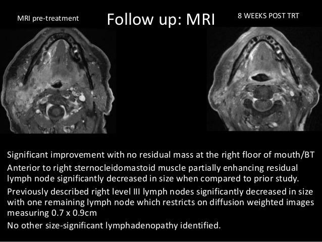
Pre And Post Pdt Mri Of Base Of Tongue Squamous Cell Carcinoma Mainly Download Scientific Diagram
Base Of The Tongue Squamous Cell Carcinoma Springerlink
Squamous Cell Carcinoma Of The Tongue Radiology Case Radiopaedia Org

Tongue Base Squamous Cell Carcinoma T2 N2b M0 Radiology Case Radiopaedia Org
Clinical Case Base Of Tongue Cancer

Base Of The Tongue Tumor Eurorad
Http Pdf Posterng Netkey At Download Index Php Module Get Pdf By Id Poster Id 137221

Tongue Squamous Cell Carcinoma Radiology Case Radiopaedia Org

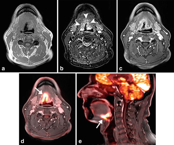
A 54 Year Old Male Patient With Right Tongue Base Oropharyngeal Download Scientific Diagram
Left Vocal Cord Lesion Squamous Cell Carcinoma Radiology Case Radiopaedia Org
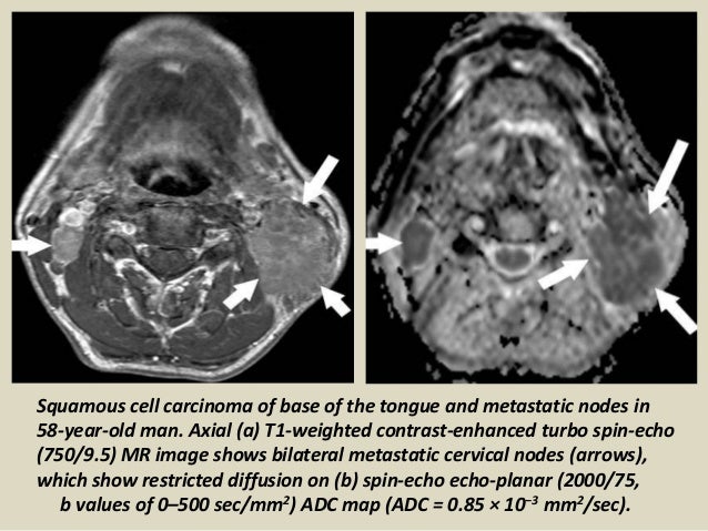
Presentation1 Radiological Application Of Diffusion Weighted Mri In

Pitfalls In The Staging Of Cancer Of The Oropharyngeal Squamous Cell Carcinoma Radiology Key

Pdf Oral Cavity Cancer
Https Encrypted Tbn0 Gstatic Com Images Q Tbn 3aand9gcrhdfaftnrpnf9bgztnywlvwvvlbgf6p8efmjbnqdnkwdbi9l9g Usqp Cau

Cancer Of The Oral Cavity And Oropharynx Abstract Europe Pmc
Primary Lesions Of The Root Of The Tongue Radiographics
Squamous Cell Carcinoma Oral Cavity Radiology Case Radiopaedia Org

Coronal T1 Mri Of Normal Oral Cavity Anatomy Ab Indicates Anterior Download Scientific Diagram

Moderately Differentiated Multifocal Squamous Cell Carcinoma Tongue Download Scientific Diagram

Tongue Squamous Cell Carcinoma Radiology Case Radiopaedia Org

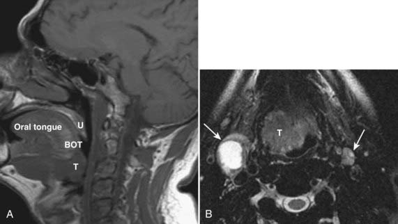
Mr Evaluation Of Tongue Carcinoma In The Assessment Of Depth Of Invasion With Histopathological Correlation A Single Center Experience Ravikanth R Indian J Radiol Imaging

Imaging The Postoperative Neck Radiology Key

Tongue Base Schwannoma Differential Diagnosis And Imaging Features With A Case Presentation Sciencedirect

Fig 3 Solid Lymph Nodes As An Imaging Biomarker For Risk Stratification In Human Papillomavirus Related Oropharyngeal Squamous Cell Carcinoma American Journal Of Neuroradiology

Oropharyngeal Squamous Cell Carcinoma Radiology Reference Article Radiopaedia Org In 2020 Squamous Cell Carcinoma Squamous Cell Squamous
Head And Neck Clinical Gate
Pitfalls In The Staging Of Cancer Of Oral Cavity Cancer Radiology Key

A 51 Year Old Patient With T3n2c Carcinoma Of The Base Of The Tongue Download Scientific Diagram
Https Pubs Rsna Org Doi Pdf 10 1148 Rg 317095738

Laryngocele A 67 Year Old Man With History Of Prior Tongue Base And Download Scientific Diagram

Left Base Of Tongue Squamous Cell Carcinoma Stage T4n2cm0 Download Scientific Diagram

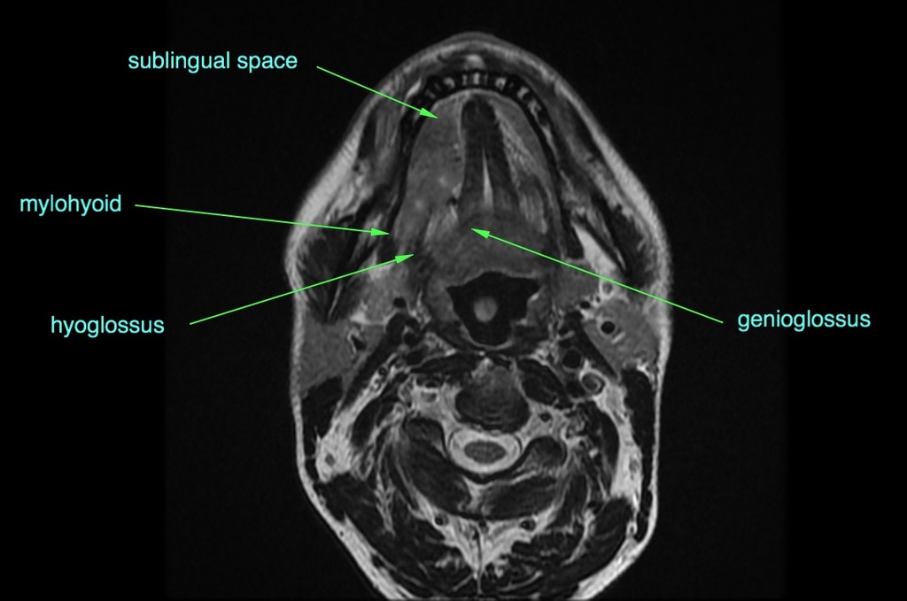
Imaging The Floor Of The Mouth And The Sublingual Space Radiographics
Https Www Iosrjournals Org Iosr Jdms Papers Vol14 Issue2 Version 5 J014253845 Pdf

Posttreatment Ct And Mr Imaging In Head And Neck Cancer What The Radiologist Needs To Know Radiographics

Oropharynx Radiology Key

Role Of Mri In Evaluation Of Malignant Lesions Of Tongue And Oral Cavity Abstract Europe Pmc

Evaluation Of Tongue Cancer Using High Resolution Sonography Dhoot 2015 Journal Of Ultrasound In Medicine Wiley Online Library

Squamous Cell Carcinoma Of The Retomolar Tigone Radiology Key
Https Encrypted Tbn0 Gstatic Com Images Q Tbn 3aand9gcqtvsdcrxvvtr 1h31c2x J 36ig9l6fz Minluaetvax 8o2ak Usqp Cau
Source : pinterest.com
